6 things you should know about plantar fasciitis
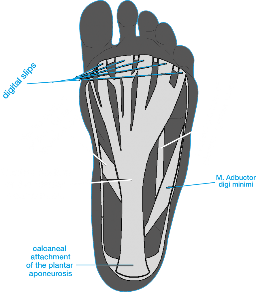
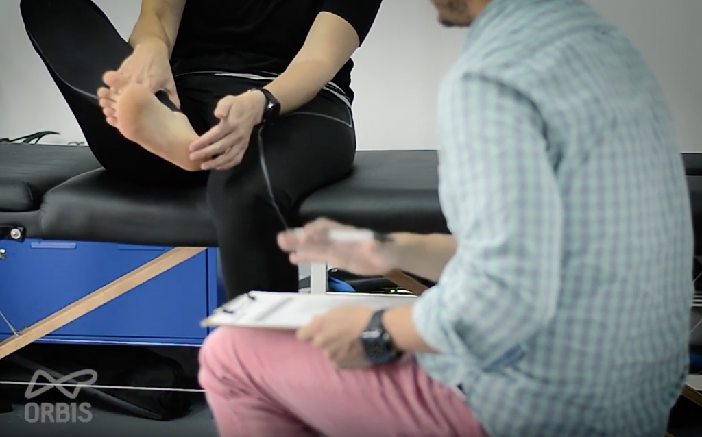
Facebook
Twitter
Email

Summary

Plantar fasciitis (PF) is an overuse injury characterised by repeated trauma to the plantar fascia, usually at the origin, the medial calcaneus tuberosity. In this post we review the plantar fascia, its function, causes of injury to this tissue and treatment options.

Nuno Gama, PhD

Nuno Gama, PhD

Nuno is passionate about science, sports, his family
and dogs. He currently rides a Fuji one.1 equipped
with Ultegra and Mavic Cosmic carbon pro.

ORBIS LaB Plantar fasciitis thumbnail

Introduction

Plantar fasciitis (PF) is the most prevalent cause of inferior heel pain (Al-Boloushi, López-Royo, Arian, Gómez-Trullén, & Herrero, 2019; Mario Roxas, 2005; Thing, Maruthappu, & Rogers, 2012) but is mainly associated with overweight individuals or runners (Cole, Seto, & Gazewood, 2005; Wilk, Fisher, & Gutierrez, 2000). Cycling is frequently prescribed as a complementary exercise in the rehabilitation of the plantar fascia (Cole et al., 2005; Mario Roxas, 2005; Thing et al., 2012; Young, Rutherford, & Niedfeldt, 2001). There is currently little evidence directly linking plantar fasciitis to the sport of cycling, however, with the rising of popularity in triathlon, many bike fitters and biomechanic scientists will have to be aware of this condition and adapt their practices to account for the influence of the high impact nature of running in cycling.

Thing 1 - What is the plantar fascia?

The plantar fascia is simply a thickened fibrous aponeurosis. An aponeurosis is simply a connective fibrous tissue similar to tendons however, in flat muscles or in flat surfaces (like the plantar of your foot), tendons are replaced by wide band with a wide area of attachment. That wide band is an aponeurosis, so if you hear someone talking about the plantar aponeurosis, you know it’s the plantar fascia they mean.

The plantar fascia has 3 bands (lateral medial and central) but it is only the central portion that originates from the medial calcaneal tuberosity on the undersurface of the calcaneus, or in the inferior heel region

ORBIS figure Anatomy of the foot showing the plantar surface.
Anatomy of the foot showing the plantar surface with the three branches of the plantar fascia

Thing 2 - What is the function of the plantar fascia?

The plantar fascia has physiological functions (Ricci, 2015) like protecting the large network of plantar blood vessels and aid in the venous return of the blood to the heart (which has to overcome the hydrostatic pressure given by the distance from the foot to the heart – aiding the foot venomuscular pumps (VMP), in other words). However, the plantar fascia also has a biomechanical function usually described by the Windlass mechanism which is a load sharing system mediated mainly by the medial longitudinal arch of the foot.
More on the Windlass mechanism here.
ORBIS LaB SERVICES

Bike fit service

Saddle fit service

Foot clinic

Lactate profile

Thing 3 - What exactly is plantar fasciitis

Sometimes the plantar fascia suffers microtears which will then trigger an inflammatory reparative response. A normal fascia has a thickness of 3 mm, but in a fasciitis it can reach 15mm of thickness. This inflammatory response in the plantar fascia is a fasciitis.

Check out our video on plantar fasciitis, below:

Thing 4 - what makes the fascia prone to tearing?

The plantar fascia can tear due to several factors such as:

– limited range of motion for ankle dorsiflexion,
– obesity,
– work related weight bearing,
– calcaneal spurs,
– fat pad atrophy.

Moreover, it is important to understand that not only athletic populations develop this condition. For example, fat pad atrophy which is the reduction of the thickness of the adipose tissue at the bottom of the heel, increases at the age of 40, which will then expose the fascia to more biomechanical stress.
Furthermore, a change in work habits, such as spending more hours standing or walking, can aggravate symptoms or facilitate the development of the condition. 

Plantar aponeurosis medial view

Thing 5 - how is plantar fasciitis diagnosed?

Diagnose of plantar fasciitis is made with a reasonable level of certainty using clinical assessment tests. The symptom most patients will report is a sharp pain localised at the bottom of the heel which is more painful with the first steps in the morning or after long periods of rest or inactivity.
Patients also report that the heel pain reduces with increasing levels of activity but will worsen towards the end of the day. History taking is important because usually there has been a change in the volume of the physical activity or simply life changes that require more hours standing or walking.
Adding to the symptoms and patient history, there are physical examinations that can help with the diagnosis, such as:
– palpation of the proximal plantar fascia insertion,
– active and passive talocrural joint dorsiflexion,
– range of motion,
– the tarsal tunnel syndrome test,
– the windlass test, and
– the longitudinal arch angle.
Clinical history taking - plantar fasciitis

Thing 6 - treatment options for plantar fasciitis

There are two type of interventions for plantar fasciitis, conservative or non-invasive and surgical or invasive. Non-invasive options are treatments such as orthoses (heels pads, plantar fascia groove, arch support), shock wave therapy, oral anti-inflammatory drugs, physiotherapy exercises, deep tissue massage, night splints, functional taping and steroid injections.

Invasive treatment options involves fascia release surgery whereby part of the fascia is cut from the calcaneus insertion to release tension. Surgery is a last resource solution and patients must be symptomatic for 12 to 24 months without relief with conventional methods.

Check out the 3 most common mistakes people recovering from plantar fasciitis make.

References

Al-Boloushi, Z., López-Royo, M. P., Arian, M., Gómez-Trullén, E. M., & Herrero, P. (2019). Minimally invasive non-surgical management of plantar fasciitis: A systematic review. Journal of Bodywork and Movement Therapies, 23(1), 122–137. https://doi.org/10.1016/j.jbmt.2018.05.002

Cole, C., Seto, C., & Gazewood, J. (2005). Plantar fasciitis: Evidence-based review of diagnosis and therapy. American Family Physician, 72(11), 2237–2242.

Mario Roxas. (2005). Plantar fasciitis: diagnosis and therapeutic considerations. Alternative Medicine Review, 10(2), 83–93.

Ricci, S. (2015). The venous system of the foot: anatomy, physiology and clinical aspects. Phlebolymphology, 22(2).

Thing, J., Maruthappu, M., & Rogers, J. (2012). Diagnosis and Management of Plantar Fasciitis in Primary Care. British Journal of General Practice, 62, 443–444. https://doi.org/10.3399/bjgp12X653769

Wilk, B. R., Fisher, K. L., & Gutierrez, W. (2000). Defective Running Shoes as a Contributing Factor in Plantar Fasciitis in a Triathlete. Journal of Orthopaedic & Sports Physical Therapy, 30(1), 21–31. https://doi.org/10.2519/jospt.2000.30.1.21

Young, C. C., Rutherford, D. S., & Niedfeldt, M. W. (2001). Treatment of plantar fasciitis. American Family Physician, 63(3), 467–474.

About the author

Nuno Gama, PhD

Nuno is the founder of ORBIS LaB, a laboratory aimed at athletes and sports enthusiasts, based in Glasgow, UK. He is an expert in biomechanics and physiology and is an extremely approachable person. Nuno enjoys talking about science, life, and sports. Nuno is passionate about his family, his dogs, his guitars and his Rubiks’ cubes.

Nuno Gama, PhD and dog cycling
Facebook
Twitter
Email

Related reading

ORBIS LaB SERVICES

Bike fit service

Saddle fit service

Foot clinic

Lactate profile